Медицина
Различные книги в жанре МедицинаPatient Blood Management
Patient Blood Management (PBM) is an innovative clinical concept that aims to reduce the need forallogenic blood transfusions, cut health-care costs, and avert or correct the risk factors related toblood transfusion, thus minimizing the rate of side effects and complications. This comprehensivehands-on volume offers a three-point approach for the implementation of PBM to improve patientoutcome, focusing on how to prevent or treat anemia, reduce blood loss, and increase anemiatolerance. The book also goes beyond preoperative PBM, with detailed accounts of coagulationdisorder management and the administration of coagulation products and platelet concentrates.Special Features:Presents a clear three-pillar strategy for the application of PBM: diagnosis and treatment of anemia, reduction of peri-interventional blood loss, and optimization of the tolerance to anemia in the everyday clinical settingCovers issues such as PBM during surgery, requirements for modern transfusion medicine, ordering blood products, the role of pre-anesthesia clinics, benchmarking processes, and potential implications of PBM in the public health sectorOverview of research in PBM including landmark studies and current clinical trialsBoxes in each chapter highlighting key information, core statements, and summariesA multidisciplinary and international team of contributors experienced in PBMPatient Blood Management is a guide for clinicians and residents whose patients are at risk for anemia, coagulation disorders, or severe blood loss. Anesthesiologists, surgeons, and specialists involved in the use of blood and blood products can use the book for quick reference or to learn more about a leading-edge concept for optimizing patient safety and improving outcome.
Abdominal Ultrasound: Step by Step
That straight-forward approach, coupled with more than 1,000 high-quality images and illustrations, enables hand-on learning, yielding the ability to assimilate these techniques quickly and adeptly. This is a stellar resource that provides the requisite tools… – Doody's Review (starred review) This third edition of a well-established text from Berthold Block maintains the same style and clarity as previous editions, taking the novice ultrasound practitioner through a series of steps across systems and levels towards scanning competency and evaluation of the findings. – RAD Magazine The third edition of this practical reference guide has been updated with a modern, visually attractive design and expanded content. The book is ideal for healthcare professionals with little or no experience in administering and interpreting abdominal ultrasound examinations. It is practice-oriented and structured in a way that allows readers with varying degrees of ultra-sonography knowledge to utilize the material according to their individual experience and needs. Each chapter includes a systematic, detailed description of the anatomy involved in the ultrasound examination, with easy-to-digest steps that follow standardized routine and protocol. That straight-forward approach, coupled with more than 1,000 high-quality images and illustrations, enables hands-on learning, yielding the ability to assimilate these techniques quickly and adeptly. This is a stellar resource that provides the requisite tools to locate and display the anatomical structure being tested, position and move the transducers accurately, describe and interpret the findings correctly, and differentiate key findings from the many image artifacts that typically occur. Key Highlights: In-depth discussion of organ boundaries, organ details, anatomical relationships, potentially abnormal findings, tips, and clearly defined learning objectives Anatomical drawings incorporate a «sliced 3-D» view that show how the structures are displayed by the sector-shaper beam Each chapter includes a series of images replicating the 3-D impression that results from the transducer moving across the body Schematic drawings illustrate the ultrasound images, including a body marker that shows the transducer position The «sono-consultant»: a systematic guide to evaluating ultrasound findings and establishing a differential diagnosis This step-by-step guide is an invaluable, pragmatic resource to have on hand while performing abdominal ultrasound on the patient. In-depth but concise, this is an essential teaching guide for medical students, residents, technicians, and physicians who need to learn and master these examination techniques.
Ophthalmology
This larger-format third edition of a remarkable atlas reflects the latest advances in the constantly evolving specialty of ophthalmology. Firsthand knowledge is culled from the authors’ decades of patient consultations, teaching, and combined surgical experience. Cataract, diabetic retinopathy, glaucoma, and age-related macular degeneration are covered along with other eye diseases affecting millions of people worldwide. Disorders less frequently seen in clinical practice are included in its comprehensive coverage.Organized anatomically, the comprehensive, this expertly written text covers the entire scope of ophthalmologic practice. Its systematic didactic outline detailing symptoms, examination findings, and diagnosis makes it easy to follow and consult.Key Features:A detailed chapter on clinical ophthalmological examinationAnatomical, pathophysiological, diagnostic, and clinical data on every area of the eye600 highest quality color photographs, superb illustrations, and overviews illustrate clinical findings and pathophysiologyTables include important medications, reference dimensions, and standard values, cardinal symptoms and diagnosesA glossary of technical ophthalmological termsNew design and larger, clearer formatMedical students and ophthalmology residents will find this an invaluable reference tool. This concise compendium also offers a stellar study aid for board exam preparation, and provides an excellent ophthalmology refresher course for practicing clinicians and allied healthcare professionals.
Functional Anatomy for Physical Therapists
Effective examination and treatment in physical therapy rely on a solid understanding of the dynamics of the joints and the functions of the surrounding muscles. This concise instructional manual helps readers to not only memorize anatomy but also to truly comprehend the structures and functions of the whole body: the intervertebral disk, the cervical spine, the cranium, the thoracic spine, the thorax, the upper extremities, lumbar spine, pelvis and hip joint, and the lower extremities. Through precise descriptions, efficiently organized chapters, and beautiful illustrations, this book relates functional anatomy to therapy practice. It provides extensive coverage of the palpation of structures and references to pathology throughout. Highlights: Accurate and detailed descriptions of each joint structure in the body, including their vessels and nerves, and their function Comprehensive guidance on the palpation of individual structures Detailed discussions on the functional aspects of muscles and joint surfaces, and the formation of joints Concise tips and references to pathology to assist with everyday practice More than 1000 illustrations clearly depicting anatomy and the interconnections between structures Physical therapists will find Functional Anatomy for Physical Therapists invaluable to their study or practice. It makes functional anatomy easier for students to learn and is ideal for use in exam preparation. Experienced therapists will benefit from practical tips and guidance for applying and refining their techniques.
Vasculature of the Brain and Cranial Base
Four master neurosurgeons bring a wealth of collective neurosurgical and neuroendovascular experience to this remarkable reference book, which melds a detailed anatomical atlas with clinical applications. The authors provide case reviews and pearls that demonstrate how anatomy impacts clinical practice decisions for aneurysm, stroke, and skull-base disease.Highlights:Comprehensive variations of the vasculature at the Circle of Willis, cortical branches, and secondary arteriesRange and average measurements of the most critical vesselsHundreds of color photographs elucidate precise anatomical cadaver dissections Exquisite illustrations by Paul H. DresselThis richly illustrated, comprehensive anatomical resource is a must have for neurosurgeons, neuroradiologists, and neurologists. Whether you are a practicing clinician or resident, reading this book will greatly expand your «vision» and sharpen your perception.
Atlas of Peripheral Regional Anesthesia
Praise for the previous edition:"This unique book encompasses everything from hearing science and psychoacoustics to hearing conservation and basic audiometry…explaining it at beginner's level while providing a more in-depth look for the more experienced." – Doody's Review Now in a more user-friendly format, with a four-color design, this new edition includes the latest scientific and clinical knowledge to give audiology students a solid understanding of core audiologic concepts. Every essential topic in audiology, from acoustics and anatomy to auditory disorders and hearing loss, is covered in this book.Key Features of the Fourth Edition:Covers new technology for electrophysiological assessment as well as bone-anchored hearing aids and cochlear implantsExpanded discussion of management techniques, now in two separate chaptersMore than 300 exquisite full-color illustrationsQuestions and answers at the end of each chapter for study and review of essential topicsExtensive bibliography with references to current literatureEssentials of Audiology, Fourth Edition , is an indispensable reference for undergraduate and first year graduate students in audiology as well as a valuable resource for speech and language pathology students. With thorough coverage of the essentials of clinical practice, this new edition is also a good refresher for audiologists and speech-language pathologists who are starting out in their practice.
Surgery for Cochlear and Other Auditory Implants
Hearing loss is one of the leading contributors to the global disease burden. In particular, the increasing population of people aged 65 years and older is expected to be a key driver. In children, hearing screening programs are becoming a worldwide standard, improving auditory rehabilitation options from a very young age. Implantable auditory technology–which, apart from cochlear implants, includes auditory brainstem implants, bone anchored hearing aids, and implantable middle ear devices–is an emerging field, and these devices represent a new era in hearing rehabilitation. However, cochlear implantation is still the biggest segment in the hearing implants market. Cochlear implantation is performed in a growing number of patients worldwide, and the indications are widening, helped by technological advances. This comprehensive, high-level surgical reference and atlas is tailored for surgeons who are undertaking training for cochlear implant procedures and implantable auditory devices and for experienced surgeons who would like to expand their knowledge, improve their skills and outcomes, and learn advanced surgical techniques. Following the principle underlying Professor Sanna's other successful publications, Surgery for Cochlear and Other Auditory Implants takes an integrated approach to anatomy, imaging, technology, decision making, surgical procedures described step by step, and clinical cases. This allows readers to:Improve the efficiency and outcomes of cochlear implantation and other auditory implant surgeriesLearn the required basic and advanced surgical techniquesEvaluate different surgical options and types of implantsReview common and uncommon variations of anatomy and malformationsUnderstand issues and surgical modifications unique to pediatric cochlear implantation, to revision surgery, and in postmeningitis, otosclerosis, and NF2 casesFind decision-making algorithms for difficult pathologiesExamine common and not so common intraoperative dilemmas and identify strategies to resolve themReview preoperative assessment and set up and outcomesFind out about classification systems in cochlear implant failure, malformations, otosclerosis, and post meningitis Supplementing the 1200 images within the book are 15 outstanding videos available on Thieme's MediaCenter demonstrating the implantation of the different cochlear implantation devices that are currently available and the application of brainstem implants in these situations: tumor removal, malformation (missing auditory nerve in children), and cochlear ossification.
Atlas of Breast Surgery
The Atlas of Breast Surgery presents the anatomy, diagnostic procedures, and step-by-step guidelines necessary for plastic surgeons, oncologists, and general surgeons to correctly and successfully perform surgical procedures on the breast. This excellent clinical reference is a superb work of art with stunning drawings and systematic tabular presentations that enhance the text. The clear and uncomplicated design will help surgeons master procedures quickly and efficiently. Key Features: More than 200 outstanding high-quality watercolor drawings Edited by internationally renowned experts on breast surgery Modern approach, emphasizing breast-conserving strategies where oncologically feasible and in the best interests of patients Detailed, point-by-point descriptions of routine as well as more complex surgical procedures Comprehensive coverage of esthetic and reconstructive techniques as well as essential procedures for the diseased breast Plastic surgeons, oncologists, and general surgeons treating patients indicated for breast surgery will undoubtedly want to refer to this atlas repeatedly in the course of their daily practice, and it is sure to become a treasured volume in their medical library for many years. As such, it constitutes a worthy and fitting companion volume to Thieme’s celebrated Atlas of Gynecologic Surgery, now in its 4th edition.
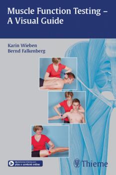
Muscle Function Testing - A Visual Guide
This beautifully illustrated pocket atlas provides physical therapists, occupational therapists, sports therapists, and students with practical guidelines and quick tests for evaluating gross motor function throughout the body. The tests in this manual are particularly suitable for analyzing isolated muscle deficits and evaluating other testing methods. When used as a regular part of the physical therapy routine, manual muscle testing provides valuable information on individual treatment needs, enables the therapist to monitor progress and modify procedures, and allows the patient to see the results for themselves.Key features: Almost 200 high-quality color photographs and illustrations help demonstrate each step in the testing processQuick tests for evaluating overall muscle function, followed by detailed guidelines for testing muscle function in the head and face, spine, and upper/lower extremitiesDetailed introductory chapter on the foundations and anatomical basis of muscle testingClear descriptions of clinical symptoms for each muscle group, plus examples from practiceOnline access to assessment forms on Thiemes MediaCenterTest questions and answers for self-studyThis book is a valuable resource for all PT practitioners and students that will enrich their practice and help them to successfully evaluate and treat patients suffering from muscle-related injuries.


